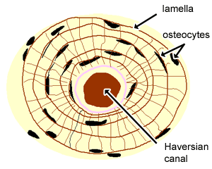
Cross Section Of A Compact Bone : Bone Structure Anatomy And Physiology I / Compact bone is very different from the other tissues you have seen.. Lamellae are found filling the spaces between circular osteons and. It is dense (because of calcified matrix) with tiny spaces known as lucanas. Within each lamella, collagen is mixed with inorganic minerals like magnesium, calcium and phosphorus and layered around a haversian canal. Cross section of a compact bone. By printing out this quiz and taking it with pen and paper creates for a good variation to only playing it online.
Transverse section of compact bone on examining a cross section of any bone, it is composed of two kinds of bony tissue: To the left is muscle tissue, and to the right In long bones, as you move from the outer cortical compact bone to. Due to the strong nature of compact bone, compared to spongy bone, it is the preferred tissue for strength. Smooth muscle fibers teased apart 1.
Cartilage Bone Ossification The Histology Guide from www.histology.leeds.ac.uk Their presence in your section will depend on the area of bone from which your section was taken. A diagrammatic view of a cross section of bone. ( ) each osteon has a central haversian canal , running parallel to long axis of bone. A cross section of a compact bone shows concentric circles called lamellae. A cross section of a compact bone shows concentric circles called lamellae. 1.compact and spongy bones are the two main types of osseous tissues. Circumferential lamellae lie along the inner and outer margins of compact bone, running along the inner and outer circumferences of the bone organ, respectively. It needs to be very strong as it supports your body and muscles as you walk, run, and move throughout the day.
Because of its strength, the compact bone makes it possible for the bone to support weight.
The cross section of this circular cylinder is a circle. Smooth muscle fibers teased apart 2. The little black spots are osteocytes. Their presence in your section will depend on the area of bone from which your section was taken. Compact bone decalcified cross section. In long bones, as you move from the outer cortical compact bone to. Compact bone model labeled 12 photos of the compact bone model labeled compact bone labeled slide, compact bone labeling game, compact bone labeling quiz, compact bone model labeled, bone, compact bone labeled slide, compact bone labeling game, compact bone labeling quiz, compact bone model labeled Obtain a demineralized compact bone preparation (in cross section), preferably from the diaphysis of a long bone, and prepare to examine it microscopically. Lamellae are found filling the spaces between circular osteons and. Compact cross section human, ground bone, 162 x. 1.compact and spongy bones are the two main types of osseous tissues. An outer 'fibrous layer' containing mainly fibroblasts, and an inner 'cambium layer' containing progenitor cells. Über 7 millionen englischsprachige bücher.
( ) each osteon has a central haversian canal , running parallel to long axis of bone. To the left is muscle tissue, and to the right Compact bone is laid in such a manner that there are histological units seen in cross section. In long bones, as you move from the outer cortical compact bone to the inner medullary cavity, the bone transitions to spongy bone. The cross section of this circular cylinder is a circle.
Lab Examining A Long Bone from s3.studylib.net Smooth muscle fibers teased apart 1. We would like to show you a description here but the site won't allow us. Compact bone, as opposed to spongy bone, is made of cylindrical units, called osteons, that are tightly formed together. Compact bone decalcified cross section. Before placing your slide on the microscope stage, remember to read the label, examine the slide with your eye and note any visible macroscopic features that might help your examination. The remainder is cancellous bone, which has a spongelike appearance with numerous large spaces and is found in the. Transverse section of compact bone on examining a cross section of any bone, it is composed of two kinds of bony tissue: Compact bone is laid in such a manner that there are histological units seen in cross section.
This is known as the periosteum.
A cross section of a compact bone shows concentric circles called lamellae. Compact bone is the heaviest, hardest type of bone. A diagrammatic view of a cross section of bone. Compact cross section human, ground bone, 162 x. Compact bone, also called cortical bone, dense bone in which the bony matrix is solidly filled with organic ground substance and inorganic salts, leaving only tiny spaces (lacunae) that contain the osteocytes, or bone cells.compact bone makes up 80 percent of the human skeleton; ( ) each osteon has a central haversian canal , running parallel to long axis of bone. Transverse section of compact bone on examining a cross section of any bone, it is composed of two kinds of bony tissue: Smooth muscle fibers teased apart 2. We will view circumferential lamellae when we study a decalcified compact bone specimen. Compact bone is the outer layer and the spongy bone forms the inner layer. An outer 'fibrous layer' containing mainly fibroblasts, and an inner 'cambium layer' containing progenitor cells. It is dense (because of calcified matrix) with tiny spaces known as lucanas. 1.compact and spongy bones are the two main types of osseous tissues.
A central tube called a haversian canal typically runs in the same path as the length of the bone. A cross section of a compact bone shows concentric circles called lamellae. The little black spots are osteocytes. A diagrammatic view of a cross section of bone. They fill the inner layer of most bones such as the vertebrae.
Structure Of Bones Biology For Majors Ii from s3-us-west-2.amazonaws.com An outer 'fibrous layer' containing mainly fibroblasts, and an inner 'cambium layer' containing progenitor cells. Related posts of cross section of a long bone compact bone model labeled. This is known as the periosteum. Compact bone is the outer layer and the spongy bone forms the inner layer. Compact bone is the heaviest, hardest type of bone. Bone decalcification is the removal of the mineral component using an acid, leaving the bone soft and easy to cut. This slide contained a cross section of a very small bone, and you are looking at the entire thickness of the shaft of the bone. ( ) each osteon has a central haversian canal , running parallel to long axis of bone.
Compact bone cross section courtesy:
Obtain a demineralized compact bone preparation (in cross section), preferably from the diaphysis of a long bone, and prepare to examine it microscopically. The cross section of this circular cylinder is a circle. A cross section of a compact bone shows concentric circles called lamellae. Über 7 millionen englischsprachige bücher. A diagrammatic view of a cross section of bone. Decalcified compact bone looks completely different than compact bone that still has calcium salts in its matrix. The little black spots are osteocytes. Their presence in your section will depend on the area of bone from which your section was taken. Fibrous astrocytes in white matter of the brain. By printing out this quiz and taking it with pen and paper creates for a good variation to only playing it online. A cross section of a compact bone shows concentric circles called lamellae. Skull bone is a flat bone. A cross section of a compact bone shows concentric circles called lamellae.
The cross section of this circular cylinder is a circle cross section of a bone. 1.compact and spongy bones are the two main types of osseous tissues.


0 Komentar